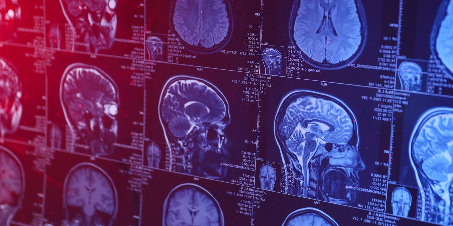
A large brain imaging study indicates that subtle changes in brain structure among antidepressant users are primarily linked to the severity of their depression rather than the medications themselves. This research, published in *Molecular Psychiatry*, also reveals that the relationship between depression, medication, and brain anatomy varies with age, showing distinct patterns in younger patients.
Read More
Major depressive disorder, a significant cause of global disability, is frequently treated with antidepressants, although the exact biological mechanisms of these medications are not fully understood. A theoretical framework established in 2006 suggested that chronic stress and depression may damage brain cells, with antidepressants potentially promoting new cell growth in deep brain regions.
Previous studies, including a 2015 report from the ENIGMA consortium—which examines how depression affects brain structure—found that individuals with major depression typically have a smaller hippocampus, a region crucial for memory and emotion.
To further investigate the effects of antidepressants on brain structure, researchers analyzed data from 32 international cohorts, involving 8,696 participants: 2,076 individuals with major depressive disorder on antidepressants, 1,495 not on medication, and 5,125 healthy controls. Lianne Schmaal, head of Mood & Anxiety Disorders Research at Orygen, emphasized the importance of the study’s scale in identifying subtle patterns in brain structure.
Participants underwent structural magnetic resonance imaging (MRI), measuring cortical thickness, surface area, and volume of subcortical structures. Statistical models adjusted for age, sex, and head size.
Findings revealed complex relationships between age, medication status, and brain structure. Younger patients on medication showed a thinner middle temporal gyrus compared to unmedicated patients and healthy controls, while this difference did not appear in older individuals. Schmaal noted that the influence of antidepressant use on brain structure varies across different age groups.
Overall, those currently on antidepressants had a smaller hippocampus and a thinner inferior temporal gyrus compared to their unmedicated counterparts. However, after adjusting for the severity of depressive symptoms, these structural differences diminished, suggesting that the observed changes could be more closely tied to the severity of depression rather than direct effects of medication.
Despite finding small differences in brain structure, researchers caution against assuming that antidepressants negatively impact brain health. Schmaal clarified that while there are observable differences, they are not significant enough to discern medication effects in individual scans. Roland Zahn, a professor at King’s College London, echoed this sentiment, stressing that factors such as illness severity must be considered when interpreting these results.
The study also found that younger individuals with depression exhibited a smaller thalamus regardless of medication use. Structural differences in brain anatomy were highlighted, where patients taking mirtazapine showed thicker regions in the brain compared to those on other antidepressants.
While the study adds significant weight to the understanding of antidepressant effects on brain structure, it remains observational. The authors highlighted the need for longitudinal research to track changes over time and understand how antidepressants influence brain development and health outcomes. Schmaal concluded that future research should clarify the relationship between antidepressant treatment and brain structure to better inform clinical practices.